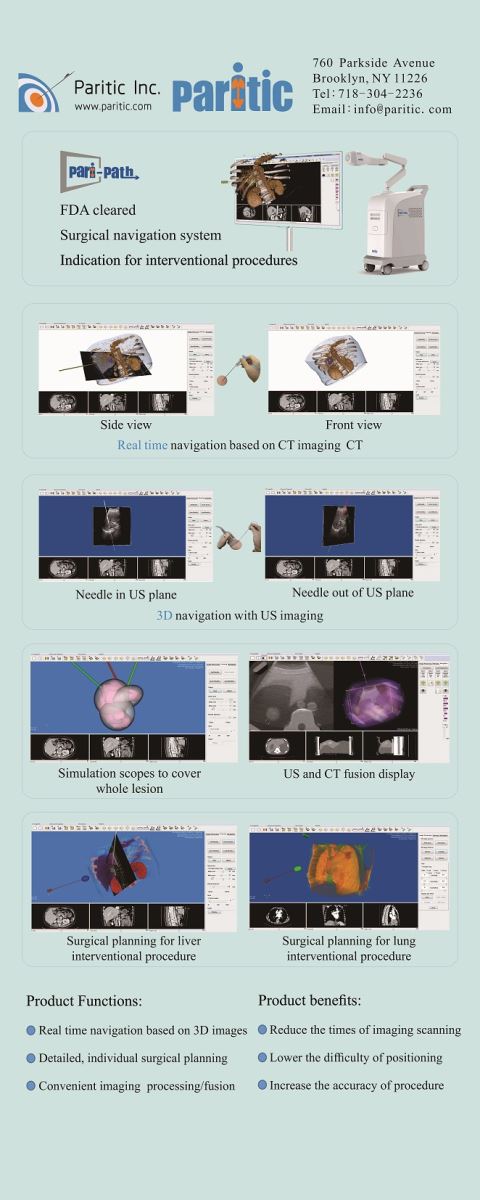
The Pari-Path surgical navigation system is a stereotactic accessory for Computed Tomography (CT) and Ultrasound imaging systems.
|
|
Pari-path surgical navigation |
Other surgical navigation |
|
Position accuracy |
about 1~2 mm* in a wide region |
In a region close to sensor |
|
Direction accuracy |
about 0.50~10 * in a wide region |
N/A |
|
Needle |
popular needle-liked instruments |
special needles only |
|
Sensor |
reusable |
expensive, disposal |
|
Register method
(mapping image with world space) |
pari-pathTM method 1. accurate in a wide region 2. register accuracy <1 mm* displayed on screen 3. no need surgeons to handle markers just a mouse click
|
metal-markers method 1.accurate in a wide region 2.need to handle markers |
|
sensors as markers 1. good accurate in a region near to sensors 2. poor accurate in a region far from sensors 3. no need to handle markers |
||
|
Fusion of CT & US method |
1. robust accuracy about 1~2 mm* 2. no need surgeons to select common spots on images |
1. accuracy not robust, maybe poor to ~cm 2. need surgeons to select common spots on images |
*Parameters (in root mean square) are typical on static objects with normal operations, may vary and are not guaranteed.
Operation steps:
|
Step ID |
Use par-pathTM navigation system |
Time (min) |
Without navigation system |
Time (min) |
|
1 |
Attach localization box on the skin |
about 1 |
Attach localization metal grids on the skin |
about 1 |
|
2 |
CT scanning |
about 3 |
CT scanning |
about 3 |
|
3 |
Transfer CT images to the navigation system via local network |
about 0.3 |
N/A |
|
|
4 |
Software operation: segment, route plan and register. optional operation: connect ultrasound, fuse CT and ultrasound |
about 2 |
Software operation on CT system: measure,route plan |
about 1 |
|
5 |
Attach reusable sensor clip to the needle |
about 1 |
N/A |
|
|
6 |
N/A
|
|
Measure and mark an inserting point on the skin surface |
about 1 |
|
7 |
Advance needle: few times |
Advance needle: multiple times |
 Click to play video.